Search This Blog

Monday, November 20, 2017

Diseases Caused by Epstein Barr Virus

Epstein barr virus is a DNA virus of family Human Herpes Virus. It is also known as HHV-4 or Human HerpesVirus 4. It is ubiquitously present and infects around 90% of world population generally resulting in mild to asymptomatic flu like symptoms.
Infectious Mononucleosis Nasopharyngeal Carcinoma Burkitt Lymphoma Hodgkin's Lymphoma B cell Lymphoma Gastric Carcinoma   Most AIDS defining Lymphomas  HIV associated CNS Lymphomas Primary Effusion Lymphoma (PEL) PlasmaBlastic Lymphoma (PBL) of Oral Cavity Environmental trigger for Systemic Autoimmune disease  SLE Rheumatoid Arthritis  Sjogren Syndrome

Infections

It causes infectious mononucleosis or glandular fever in the adolescents.  It is transmitted by saliva and infects the epithelium of nasopharynx and oropharynx and subsequently enters B cell to infect it. It manifests as pharyngitis, cervical lymphadenopathy, fatigue and fever.It is transmitted with kissing and hence is known as Kissing disease.

Cancers

First known virus to cause cancers to human was EBV affecting epithelial cells, mesenchymal cells and lymphocytes. It affects all immunocompromised and immunocompetent hosts or those with acquired iatrogenic immunosuppression. Gastric Adenocarcinoma, endemic  Burkitt Lymphoma, Hodgkins Lymphoma, large B cell lymphoma, Nasopharyngeal Carcinoma has been associated with HHV4.

Autoimmune diseases:


High viral EBV has been found to be associated with SLE and its flares. Similarly, high amount of  EBV infected B cells have been found in Synovium and joints of patient affected with Rheumatic Arthritis. Antibodies directed against EBV has been found in patients with Sjogren Syndrome and they have been found to have higher incidences of lymphoid malignancies like MALT, Non hodgkin's Lymphoma. 
EBV has been found as an environmental trigger in the development of various Systemic Autoimmune disease.


Lymphoproliferative diseases associated with HIV

EBV when occurs simultaneously with HIV, it predisposes to various lymphoproliferative diseases. It is mostly secondary to immunosuppression and are of B cell origin. About half of HIV defining cancers are associated with EBV like Hodgkins Lymphoma, Burkitt lymphoma, diffuse large B cell Lymphoma, peripheral T cell Lymphoma but more specific and  associated with HIV with EBV include Plasma Blastic lymphoma (PBL) of oral cavity, Primary effusion lymphoma (PEL), HIV related CNS lymphomas.

Sources:
https://www.ncbi.nlm.nih.gov/pmc/articles/PMC3766599/
https://www.ncbi.nlm.nih.gov/pmc/articles/PMC4314581/
https://www.ncbi.nlm.nih.gov/pmc/articles/PMC4346501/
http://www.ijcem.com/files/ijcem0012377.pdf

Friday, November 17, 2017

Hypertension in Community: Rule of halves

Population   Hypertensive   Diagnosed    Treated     Adequate     Inadequate    Untreated   Undiagnosed  Normotensive  Total Population  Hypertensive cases Diagnosed cases Treated cases
Hypertension in Community, the rule of halves.
Hypertension is a common public health problem. The trend of prevalence of hypertension is increasing with the development of newer technological, easy life style, less physical activities and increasing amount of processed food consumption.

Of the total population, 50% have hypertension both diagnosed and undiagnosed. Only 50% out of all hypertensive cases 50% seek medical attention. Of the total 50% of diagnosed cases, 50% take medicine and 50% do not. Of the 50% who take medicine, only 50% are adeuately treated and the rest 50 % are not.
Population   Hypertensive   Diagnosed    Treated     Adequate     Inadequate    Untreated   Undiagnosed  Normotensive  Total Population  Hypertensive cases Diagnosed cases Treated cases
Hypertension in Community, the rule of halves.

Tuesday, November 14, 2017

Anatomical Structures of Various length

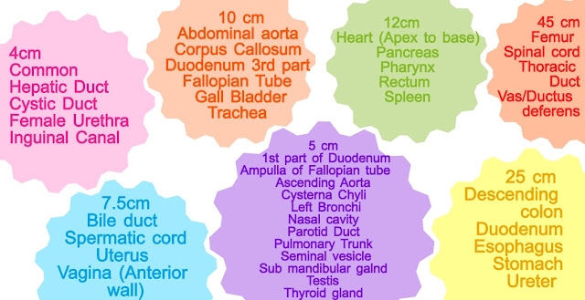
There are various structure with similar length. These are favourite question of anatomy professors. Some structures of similar lengths are given below.

45 cm structures Femur Spinal cord Thoracic Duct Vas/ Ductus deferens  25 cm Structures Descending colon Duodenum Oesophagus Stomach Ureter   12cm Structures  Heart (Apex to base) Pancreas Pharynx Rectum  Spleen  10 cm structures Abdominal aorta  Corpus Callosum  Duodenum 3rd part  Fallopian Tube  Gall Bladder Trachea    7.5cm Structure Bile duct  Spermatic cord Uterus  Vagina (Anterior wall)  5 cm Structures 1st part of Duodenum Ampulla of Fallopian tube  Ascending Aorta  Cysterna Chyli  Left Bronchi Nasal cavity  Parotid Duct  Pulmonary Trunk  Seminal vesicle Sub mandibular galnd  Testis  Thyroid glnnd   4cm Structures Common Hepatic Duct  Cystic Duct  Female Urethra Inguinal Canal


45 cm structures
Femur
Spinal cord
Thoracic Duct
Vas/ Ductus deferens

25 cm Structures
Descending colon
Duodenum
Oesophagus
Stomach
Ureter 

12cm Structures 
Heart (Apex to base)
Pancreas
Pharynx
Rectum 
Spleen

10 cm structures
Abdominal aorta 
Corpus Callosum 
Duodenum 3rd part 
Fallopian Tube 
Gall Bladder
Trachea 


7.5cm Structure
Bile duct 
Spermatic cord
Uterus 
Vagina (Anterior wall)

5 cm Structures
1st part of Duodenum
Ampulla of Fallopian tube                                  
Ascending Aorta 
Cysterna Chyli 
Left Bronchi
Nasal cavity 
Parotid Duct 
Pulmonary Trunk 
Seminal vesicle
Sub mandibular galnd 
Testis 
Thyroid glnnd 

4cm Structures
Common Hepatic Duct 
Cystic Duct 
Female Urethra
Inguinal Canal 


Thursday, November 2, 2017

List of All named Hernias (Hernia Eponyms/Special Hernia)


Hernia is the abnormal protusion of a viscous or a part of it through an opening with a sac covering it. It could either be internal or external and not necessarily from the abdominal cavity. The most commonly encountered are the inguinal hernias and femoral hernia. But there are certain hernias that are named and arise from its specific location or contain specific content.

Hernia Eponyms on the basis of their similarity with the adjacent ones

Name of Hernia
Description
Bochdalek’s Hernia
Congenital Posterolateral Hernia of Diaphargm
Morgagni Hernia4
Through Larry’s space in Diaphargm
Larry’s Hernia4
Gibbon Hernia
Hernia with Hydrocele
Bergers Hernia
Hernia in Pouch of Douglas
Romberg Hernia1
Concurrent Ipsilateral Direct and indirect Inguinal Hernia
Saddle Hernia1
Dual Hernia1
Lumbar Hernia
Hernia in lumbar triangles of type Petit’s and Grynfelt
Obturator Hernia
Hernia through Obturator Canal
Grynfelt Hernia
Upper Lumbar triangle Hernia
Petit’s Hernia
Lower Lumbar Triangle Hernia
Femoral Hernia        
Hernia medial to Femoral Vein in Femoral canal
Cooper’s Hernia6
Femoral Hernia with two sacs
Bilocular femoral Hernia6
Cloquet’s Hernia
Hernia Through Pectineal aponeurosis
Callison-Cloquet Hernia
Through Pectineus Muscle and its fascia
Narath’s Hernia
Behind Femoral Artery in congenital dislocation of Hip
Hesselbach’s Hernia
Lateral to Femoral Artery
Serofini’s Hernia
Behind Femoral Vessels
Laugier’s Hernia
Through Lacunar Ligament
Teale’s Hernia5
In front of Femoral Vessels
Velpeau Hernia5
Richter’s Hernia
Part of circumference of bowel wall is entrapped
Littre’s Hernia
Contains Meckel Diverticulum
Sliding Hernia
Hernia with a part of the wall formed by the viscus
Hernia En Glissae
Hernia with a part of the wall formed by the viscus
Maydl’s Hernia2
Two loops of bowel in Hernia sac with remaining part in abdomen
W Hernia/Hernia in W2
Retrograde Hernia2
Two loops of bowel in Hernia sac with remaining part in abdomen
Phantom Hernia
Localised Muscle Bulge following Muscle Paralysis
Mery’s Hernia7
Through the perineal floor  
Perineal Hernia7
Spigelian Hernia
Hernia through Speligian Fascia
Sciatic hernia
Hernia through greater or lesser sciatic foramen
Little’s hernia3
Appendix in inguinal  Hernial Sac
Amyand hernia3
De Garengeots Hernia
Incarceration of appendix within Femoral Hernia
Beclards Hernia
Femoral Hernia Through Saphenous Opening
Barth’s Hernia
Hernia Between Adbominal wall and Persistent Vitellointestinal Duct
Holthouse’s Hernia                   
Inguinal Hernia That has turned outward into the groin
Grubers Hernia
Internal Mesogastric Hernia
Mesocolic hernia
Transmesenteric Hernia
Kronlem Hernia
Inguinoproperitoneal Hernia
Trietz Hernia
Paraduodenal Hernia
Rieux Hernia
Retrocaecal Hernia


Hernia Eponyms in alphabetical order

Name of Hernia
Description
Amyand hernia3
Appendix in inguinal hernia Sac
Barth’s Hernia
Hernia Between Adbominal wall and Persistent Vitellointestinal Duct
Beclards Hernia
Femoral Hernia Through Saphenous Opening
Bergers Hernia
Hernia in Pouch of Douglas
Bilocular femoral Hernia6
Femoral Hernia with two sacs
Bochdalek’s Hernia
Congenital Posterolateral Hernia of Diaphargm
Callison-Cloquet Hernia
Through Pectineus Muscle and its fascia
Cloquet’s Hernia
Hernia Through Pectineal aponeurosis
Cooper’s Hernia6
Femoral Hernia with two sacs
De Garengeots Hernia
Incarceration of appendix within Femoral Hernia
Dual Hernia1
Concurrent Ipsilateral Direct and indirect Inguinal Hernia
Femoral Hernia        
Hernia medial to Femoral Vein in Femoral canal
Gibbon Hernia
Hernia with Hydrocele
Grubers Hernia
Internal Mesogastric Hernia
Grynfelt Hernia
Upper Lumbar triangle Hernia
Hernia En Glissae
Hernia with a part of the wall formed by the viscus
Hesselbach’s Hernia
Lateral to Femoral Artery
Holthouse’s Hernia                   
Inguinal Hernia That has turned outward into the groin
Kronlem Hernia
Inguinoproperitoneal Hernia
Larry’s Hernia4
Through Larry’s space in Diaphargm
Laugier’s Hernia
Through Lacunar Ligament
Little’s hernia3
Appendix in inguinal  Hernial Sac
Littre’s Hernia
Contains Meckel Diverticulum
Lumbar Hernia
Hernia in lumbar triangles of type Petit’s and Grynfelt
Maydl’s Hernia2
Two loops of bowel in Hernia sac with remaining part in abdomen
Mery’s Hernia7
Through the perineal floor
Mesocolic hernia
Transmesenteric Hernia
Morgagni Hernia4
Through Larry’s space in Diaphargm
Narath’s Hernia
Behind Femoral Artery in congenital dislocation of Hip
Obturator Hernia
Hernia through Obturator Canal
Perineal Hernia7
Through the perineal floor
Petit’s Hernia
Lower Lumbar Triangle Hernia
Phantom Hernia
Localised Muscle Bulge following Muscle Paralysis
Retrograde Hernia2
Two loops of bowel in Hernia sac with remaining part in abdomen
Richter’s Hernia
Part of circumference of bowel wall is entrapped
Rieux Hernia
Retrocaecal Hernia
Romberg Hernia1
Concurrent Ipsilateral Direct and indirect Inguinal Hernia
Saddle Hernia1
Concurrent Ipsilateral Direct and indirect Inguinal Hernia
Sciatic hernia
Hernia through greater or lesser sciatic foramen
Serofini’s Hernia
Behind Femoral Vessels
Sliding Hernia
Hernia with a part of the wall formed by the viscus
Spigelian Hernia
Hernia through Speligian Fascia
Teale’s Hernia5
In front of Femoral Vessels
Trietz Hernia
Paraduodenal Hernia
Velpeau Hernia5
In front of Femoral Vessels
W Hernia/Hernia in W2
Two loops of bowel in Hernia sac with remaining part in abdomen


The number in superscript suggest that the hernias with the same number are actually same with various names.


Sources
SRB’s Manual Of Surgery
https://medicallegends.blogspot.com/2015/08/named-hernias.html
http://lessons4medicos.blogspot.com/2009/07/different-eponymous-types-of-hernia.html
Internet