Search This Blog

Showing posts with label Lower Motor Neuron. Show all posts

Saturday, May 12, 2018

Neurogenic Bladder: UMN vs LMN: Physiology made easy.

Basic Physiology of Micturition

Micturition is a spinal reflex modulated by CNS.
Neural control of micturition.
Source: http://physiologyplus.com/micturition-reflex-steps/

The pre-frontal cortex is responsible for the cognitive control of the micturition which analyses the signals from bladder and conveys signals according to void or not to void depending on the social setting.

The higher control of micturition is mediated by pontine micturition centre (PMC) from where nerve fibre arise and travel along the lateral columns bilaterally. It is the mechanical control of micturition. It coordinates the function of baldder and sphincter.

The Sympathetic fibres are thoracolumbar (T10-L2) outflow of nerve fibres and terminate in the hypogastric ganglion.

The Parasympathetic fibres are sacral (S2-S4) outflow.

The voluntary control over the external urtheral spincter is mediated by somatic fibres of Pudendal nerve. 
Intact Spinal cord is essential for normal micturition as it serves as a intermediate relay between the brain and the sacral center of micturition. Sacral reflex center is the primitive voiding center which is responsible for infants diaper need, since there is a continuous cycle of bladder filling and voiding. The higher mental function gradually enhances in kids as they are growing and accordingly they are trained to use toilet with their enhanced higher mental function.


Sympathetic
ParaSympathetic
Bladder (Detrusor Muscle)
Relaxation
Contraction
Bladder Neck
Contarction
Relaxation

The bladder wall is relaxed and the neck constricted with sympathetic stimulation which allows for retention of urine. The parasympathetic stimulation causes bladder wall to contract and sphincter to relax easing the voiding of urine.

Analogy of Skeletal Muscle contraction and Bladder
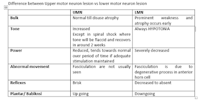
Character  UMN Type  LMN Type   Spastic Bladder Flaccid Bladder Tone  Hypertonic (Increased) Hypotonic (Decreased) Volume  Normal or small Large Detrussor contraction  Involuntary intermittent contractions (Overactiity) Absent  (Underactivity)  Pressure  High  Low Incontinence type Urge  Overflow  Symptom Urgency and Frequency  nocturia  Leaking of urine  Dribbling of urine  Erectile edysfunction in men  Retention  Incomplete bladder voiding  (Detrussor-Sphincter Dyssynergia) Uncoordinated bladder contraction and sphincter relaxation Detrusor Aflexia  Conditions  Spinal Cord damage above T12 Cerebrovascular accidents   Spinal cord damage at S2-S4 Peripheral Nerve injury  Acute Stage of spinal cord injury  Cauda Equina, Conus medullaris
LMN vs UMN Lesion : Effect on Bladder

There is a lot of analogy between skeletal muscle contraction and bladder.

In the absence of higher control, overdistension of bladder causes reflex detrusor contraction. Similar to     the muscle stretch reflex mediated by spindle fibre in skeletal muscle.

The upper motor neuron lesion of the brain and the spinal cord causes features similar to that of the UMN lesion of in the muscle characterized by Spastic bladder/ Hypertonic baldder.This is due to the reflex detrusor contraction. There is increased tone of the detrusor muscle. However, the bladder contracts with overdistension, the sphincter does not relax causing bladder sphincter dyssyenrgia. This causes urgency and urge incontinence. The volume of residual urine in the bladder is increased which causes high risk for UTI and chronic renal failure due to obstructive uropathy. The site of the lesion is generally the Spinal cord or pons or higher. There is no gross dilatation of the bladder due to the reflex contraction which results in low volume high pressure inside the bladder.


The lower motor neuron lesion to the fibre supplying bladder causes overflow incontinence. This occurs because bladder is overdistended however the reflex detrusor contraction doesnot comes into play. So what happens is the bladder leaks over time when it is beyond its holding capacity without the detrusor muscle contracting. The bladder is grossly dilated resulting in high vomule and low ressure inside the bladder.This can be described as flaccid or atonic bladder similar to flaccid paralysis of muscles in LMN lesion.The patient cannot initiate the micturition. The site of injury is generally the sacral fibres or peripheral nerve fibres

The last type of neurogenic bladder ocuurs due to injury in the prefrontal cortex which is responsible for social control of micturition. It allows us to find us to micturate in appropriate place. The patient doesnot have the sense of bladder fullness. They have trouble initiating micturition and they micturitate at inappropriate places.

Character
UMN Type
LMN Type

Spastic Bladder
Flaccid Bladder
Tone
Hypertonic (Increased)
Hypotonic (Decreased)
Volume
Normal or small
Large
Detrussor contraction
Involuntary intermittent contractions
(Overactiity)
Absent
(Underactivity)
Pressure
High
Low
Incontinence type
Urge
Overflow
Symptom
Urgency and Frequency
nocturia
Leaking of urine
Dribbling of urine
Erectile edysfunction in men
Retention
Incomplete bladder voiding
(Detrussor-Sphincter Dyssynergia)
Uncoordinated bladder contraction and sphincter relaxation
Detrusor Aflexia
Conditions
 Spinal Cord damage above T12
Cerebrovascular accidents

Spinal cord damage at S2-S4
Peripheral Nerve injury
Acute Stage of spinal cord injury
Cauda Equina, Conus medullaris
  
Source: Davidson, Merck’s Manual, Medscape
Sunday, September 3, 2017

Difference between UMN and LMN lesion: Physiology


UMN
LMN
Bulk
Normal till disuse atrophy
Prominent weakness and atrophy occurs early
Tone
Increased
Except in spinal shock where tone will be flaccid and recovers in around 2 weeks
Always HYPOTONIA
Power
Reduced, tends towards normal over period of time if adequate stimulation maintained
Severely decreased
Abnormal movement
Fasciculation are not usually seen
Fasciculation is due to degenerative process in anterior horn cell
Reflexes
Brisk
Decreased to absent

Plantar/ Babiknsi
Up going
Downgoing


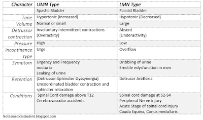
The most important thing to know is the intactness of the muscle stretch reflex. The tone is maintained under the influence of gamma motor neuron by the alpha motor neuron and the bulk and power is the pure function alpha motor neuron. The reflex is complete if its loop is complete.
alpha motor neuron , gamma motor neuron, corticospinal tract muscle spindle, extrafusal, intrafusal muscle fibre
Muscle Reflex arc
Upper Motor Neuron lesions are the lesion occurring anywhere in the central nervous system from the brain upto the spinal cord before the alpha motor neurons arise from the spinal cord. The lesion could arise from the cerebral cortex, internal capsule, midbrain, pons, medulla and the cortico spinal tract in the spinal cord. The lesions can be anything from vascular, traumatic, degenerative, and inflammatory to infective.

The lesions occurring after the alpha motor neuron accounts for the lower motor neuron lesions. The lesions could arise in the nucleus of alpha motor neuron (Polio myelitis, Amylotrophic lateral Sclerosis, brown sequard syndrome), Lesions in the nerve (Traumatic resection, entrapement, neuritis), Lesion in the NMJ (Myasthenia gravis, Lambert Eaton Syndrome) and the muscle (Duchenne Muscular Dystrophy, Beckers Muscular Dystrophy )itself.

The two principle that determine the features of upper motor and lower motor neuron include the completeness of muscle reflex arc and the higher motor control over it.
The motor reflex arc consists of the Ia fibres carrying the signal from muscle spindle which prevents the excessive stretch of muscle by contracting it. The afferent fibres mono synaptically fires the alpha motor neurons at the anterior horn and causes the muscle contraction. This reflexes is controlled further by higher centre and is thus there is an inhibitory mediatory released via an intermediate neuron coming from the corticospinal tract. If this closed loop of nerve are intact the muscle tendon reflex is intact and so is the deep tendon reflex which is elicited using reflex hammer.
The tone is the inherent state of contraction of muscles to maintain the posture of the body. It is in medicated by the Gamma motor neuron coming together wit alpha motor neuron and innervates the intrafusal fibres of muscle spindle and thus increases the sensitivity of change in length of the muscle. It is also directly innervated from the corticospinal tract and thus is affected in response to UMN lesion.  Along with that the gamma motor neurons are spontaneously firing and thus influence the sensitivity of alpha motor neurons and thus affect the tone.

In lower motor neuron lesion, the alpha motor neuron and distal is injured. So the loop can not be complete and hence no reflex contraction of muscle in response to stretch of muscle spindle receptor. In upper motor neuron lesion, the higher inhibition over the reflex arc is lost. This causes the excessive firing from alpha motor neuron and hence exaggerated deep tendon reflex.
The lower motor neurone lesion will develop flaccid paralysis because there is no innervation to muscle fibre to cause its contraction and hence they easily go into disuse atrophy early and the bulk is reduced. Contrary to the Upper motor neuron lesion where the higher control of the muscle is lost but still the muscle can be contracted locally. So, constant use of muscle via passive movement can preserve the bulk of the muscle.

The tone in upper motor neuron lesion is exaggerated because the supraspinous modulation over the gamma motor neuron is lost and they are firing spontaneously.  This increases the tone of the muscle with increases sensitivity of muscle spindle to passive stretch and increased firing in the Ia fibres. This increased firing induces increased firing in alpha motor neuron and increased contraction.The tone is higher in the antigravity muscle and hence clasp knife rigidity is due to the greater bulk of the antigravity muscles and hence the paralysis is spastic type in upper motor neuron lesion.

However, in the lower motor neuron lesion , again the same nerve that complete the muscle reflex is incomplete and hence the normal tone is present due to absence of innervation in the muscle to bring about the contraction and hence the tone is flaccid and hence the paralysis flaccid paralysis.

In LMN lesion, the muscle become hypersensitive to neurotransmitter as it is denervated. Similarly the damaged lower motor erratically discharges the neurotransmitter stored within itself as the neuron degrades. So, both increased hypersensitivity and erratic release of neurotransmitter causes fasciculations. However, in UMN lesion, there is regular firing to prevent the atrophy of muscles.

Learn about Cranial nerve examinations 

Upper Motor neuron , Lower motor neuron, differnence
Difference between UMN and  LMN lesion in tabulated form.