Search This Blog

Showing posts with label Clinical Examination. Show all posts

Saturday, May 12, 2018

Neurogenic Bladder: UMN vs LMN: Physiology made easy.

Basic Physiology of Micturition

Micturition is a spinal reflex modulated by CNS.
Neural control of micturition.
Source: http://physiologyplus.com/micturition-reflex-steps/

The pre-frontal cortex is responsible for the cognitive control of the micturition which analyses the signals from bladder and conveys signals according to void or not to void depending on the social setting.

The higher control of micturition is mediated by pontine micturition centre (PMC) from where nerve fibre arise and travel along the lateral columns bilaterally. It is the mechanical control of micturition. It coordinates the function of baldder and sphincter.

The Sympathetic fibres are thoracolumbar (T10-L2) outflow of nerve fibres and terminate in the hypogastric ganglion.

The Parasympathetic fibres are sacral (S2-S4) outflow.

The voluntary control over the external urtheral spincter is mediated by somatic fibres of Pudendal nerve. 
Intact Spinal cord is essential for normal micturition as it serves as a intermediate relay between the brain and the sacral center of micturition. Sacral reflex center is the primitive voiding center which is responsible for infants diaper need, since there is a continuous cycle of bladder filling and voiding. The higher mental function gradually enhances in kids as they are growing and accordingly they are trained to use toilet with their enhanced higher mental function.


Sympathetic
ParaSympathetic
Bladder (Detrusor Muscle)
Relaxation
Contraction
Bladder Neck
Contarction
Relaxation

The bladder wall is relaxed and the neck constricted with sympathetic stimulation which allows for retention of urine. The parasympathetic stimulation causes bladder wall to contract and sphincter to relax easing the voiding of urine.

Analogy of Skeletal Muscle contraction and Bladder
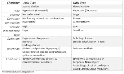
Character  UMN Type  LMN Type   Spastic Bladder Flaccid Bladder Tone  Hypertonic (Increased) Hypotonic (Decreased) Volume  Normal or small Large Detrussor contraction  Involuntary intermittent contractions (Overactiity) Absent  (Underactivity)  Pressure  High  Low Incontinence type Urge  Overflow  Symptom Urgency and Frequency  nocturia  Leaking of urine  Dribbling of urine  Erectile edysfunction in men  Retention  Incomplete bladder voiding  (Detrussor-Sphincter Dyssynergia) Uncoordinated bladder contraction and sphincter relaxation Detrusor Aflexia  Conditions  Spinal Cord damage above T12 Cerebrovascular accidents   Spinal cord damage at S2-S4 Peripheral Nerve injury  Acute Stage of spinal cord injury  Cauda Equina, Conus medullaris
LMN vs UMN Lesion : Effect on Bladder

There is a lot of analogy between skeletal muscle contraction and bladder.

In the absence of higher control, overdistension of bladder causes reflex detrusor contraction. Similar to     the muscle stretch reflex mediated by spindle fibre in skeletal muscle.

The upper motor neuron lesion of the brain and the spinal cord causes features similar to that of the UMN lesion of in the muscle characterized by Spastic bladder/ Hypertonic baldder.This is due to the reflex detrusor contraction. There is increased tone of the detrusor muscle. However, the bladder contracts with overdistension, the sphincter does not relax causing bladder sphincter dyssyenrgia. This causes urgency and urge incontinence. The volume of residual urine in the bladder is increased which causes high risk for UTI and chronic renal failure due to obstructive uropathy. The site of the lesion is generally the Spinal cord or pons or higher. There is no gross dilatation of the bladder due to the reflex contraction which results in low volume high pressure inside the bladder.


The lower motor neuron lesion to the fibre supplying bladder causes overflow incontinence. This occurs because bladder is overdistended however the reflex detrusor contraction doesnot comes into play. So what happens is the bladder leaks over time when it is beyond its holding capacity without the detrusor muscle contracting. The bladder is grossly dilated resulting in high vomule and low ressure inside the bladder.This can be described as flaccid or atonic bladder similar to flaccid paralysis of muscles in LMN lesion.The patient cannot initiate the micturition. The site of injury is generally the sacral fibres or peripheral nerve fibres

The last type of neurogenic bladder ocuurs due to injury in the prefrontal cortex which is responsible for social control of micturition. It allows us to find us to micturate in appropriate place. The patient doesnot have the sense of bladder fullness. They have trouble initiating micturition and they micturitate at inappropriate places.

Character
UMN Type
LMN Type

Spastic Bladder
Flaccid Bladder
Tone
Hypertonic (Increased)
Hypotonic (Decreased)
Volume
Normal or small
Large
Detrussor contraction
Involuntary intermittent contractions
(Overactiity)
Absent
(Underactivity)
Pressure
High
Low
Incontinence type
Urge
Overflow
Symptom
Urgency and Frequency
nocturia
Leaking of urine
Dribbling of urine
Erectile edysfunction in men
Retention
Incomplete bladder voiding
(Detrussor-Sphincter Dyssynergia)
Uncoordinated bladder contraction and sphincter relaxation
Detrusor Aflexia
Conditions
 Spinal Cord damage above T12
Cerebrovascular accidents

Spinal cord damage at S2-S4
Peripheral Nerve injury
Acute Stage of spinal cord injury
Cauda Equina, Conus medullaris
  
Source: Davidson, Merck’s Manual, Medscape
Friday, September 1, 2017

Abdominal Examination: How to describe.


Describing an abdominal examination.
Inspection
Abdomen is flat (scaphoid/distended), moving corresponding with respiration (silent).
The umbilicus is centrally (more towards xiphisternum or symphysis pubis) placed and inverted (slit like/everted).
Flanks are not full.
A horizontal scar of around 15cm around 5 cm away from the umbilicus anteriorly and 5 cm away from vertebrae posteriorly us present. Multiple erythematous papules present over the abdomen.
No visible or dilated veins, no visible peristalsis and no visible pulsations present.
No supraclavicular and renal angle fullness.
Palpation
Palpation of Liver 
On palpation, there is no local rise in temperature and no tenderness.
No masses palpable on superficial palpation and deep palpation. (If any mass palpable describe the mass accordingly in terms of size, shape, tenderness, consistency, border, margin, intraperitoneal vs retroperitoneal vs abdominal wall)
Liver and spleen were not palpable. (If organomegaly describe according to its size from anatomic landmarks, tenderness, surface, margins, borders, consistency. If liver is palpable, the percussion of the chest for its upper margin and the liver span should also be mentioned.)
Kidneys were not ballotable.
Renal angle tenderness present over the right side.
No gross deformity seen on genital examination
Supraclavicular lymph nodes were not palpable.

Percussion
Tympanic note was present all over the abdomen. (If dullness present, mention about shifting dullness and fluid thrill.)

Auscultation
Normal bowel sounds heard, 3/min.

Per rectal examination was not performed.
>>No abdominal examination is complete without digital rectal examination. 


Thursday, August 31, 2017

Examination of Ulcer: How to describe.


Examination of an Ulcer
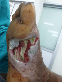
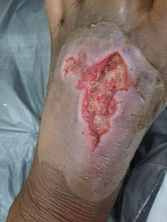
(The description below does not match with the images given beside the text. The images are only for illustrative purposes.)

Inspection

Ulcer over the medial aspect of foot extending into the sole.
Ulcer over the sole.
On inspection of the right leg, the leg is slightly elevated on the pillow and slightly flexed over the knee joint. (Attitude of the limb).

A single (number), irregularly shaped (shape) ulcer of size around 10 cm x 5cm (size) is present over the dorsum of the foot (site), extending from the lateral malleolus up to the base of the toes (extension).

The floor of the ulcer is pinkish to red (color) with minimal (amount) serous (type of discharge) discharge. No slough, foreign bodies or any other debris (Content on the floor). Multiple tendons are visible towards the distal end (base).

The edge of the ulcer is sloping type (character), with healthy pink granulation tissue (content) and the margin is sharp and regular.

The surrounding skin is exfoliated upto the level of ankle joint. The skin around the ulcer is non-erythematous, and non-edematous. No scars, abnormal pigmentation, or excoriation marks present.

The joint mobility of the proximal and distal joint along with that of the entire limb is not impaired. (Gross motor status and Range of motion of the affected joints)

Palpation
Ulcer over the dorsum of left foot.
Ulcer over the dorsum of foot

On palpation, there is local rise in temperature and mid tenderness over the ulcer.

The edges are not indurated and the base of the wound are also not indurated. The ulcer is 3-4mm deep, does not bleed on touch, and mobile over the underlying base.

The skin around the ulcer has mild rise in temperature locally with tenderness present. The skin is freely mobile on the underlying structure.

Dorsalis pedis artery, anterior tibial and posterior tibial artery are palpable and bilaterally symmetrical. Capillary refill time over the distal phalanges is more than 2 s.(Gross vascular status)

Active and passive range of motion over the limb is not restricted. Sensation is grossly intact over the area distal to the ulcer. (Gross Neurological status and range of motion)

The draining lymph nodes over the left inguinal region are not palpable significantly.


Ulcer examination is incomplete without draining lymph nodes palpation. 


Digital Rectal Examination: How to describe

Describing a rectal examination and prostate

(Per Rectal examination is a part of abdominal examination and no per rectal examination is complete without perineal examination, per rectal examination and examination of supraclavicular node (Nodes of Virchow) along with renal angle tenderness. The following describes a normal finding of per rectal examination. If any abnormality or significant findings it must be described 
accordingly.)

Inspection
On inspection of the perineal area, no redness, no any perianal mass or tag, no fissure, no ulceration or any sinus opening seen.

Palpation
No palpation of the perineal area, there is no local rise in temperature, no tenderness felt. No any palpable mass present.

Digital Rectal Examination
The tone of the sphincter is normal with no tenderness is present.
The anal canal is empty and no hard masses, impacted fecal material and polyps are palpable.
The mucosa overlying the anal canal is soft and mobile. There is no any palpable ulceration and fungating mass on examination of the walls of anal canal.
The prostate is enlarged and the upper border can not be palpated. The consistency is firm, the surface is smooth and borders are regular. The median sulcus can be palpated well and the mucosa overlying the prostate is freely mobile.
No blood stain was present on the tip of the finger at the end of digital Rectal Examination.


Wednesday, July 5, 2017

Complete Chest Examination (Respiratory System): How to describe.


>>May be first you want to see how to write a history of COPD.

Elderly  gentleman, ill looking, thin built (with Temporal hollowing, Buccal Hollowing, Supraclavicular, suprasternal hollowing, Subclavicular hollowing, with thin limbs, thin skin and prominently visible vessels) lying in semirecumbent position with IV canula of 20G and nasal prongs delivering 3l of O2/min is conscious, cooperative and well oriented to time, place and person.

On general physical examination, there is central cyanosis with peripheral cyanosis.
Bilateral pitting edema extending upto the mid thigh is present which is non tender with normal overlying skin. Sacral edema is present.
But no clubbing, no pallor, no icterus seen. Hydration status of the patient is normal. Accessible Lymph nodes are not palpable.

On examination of the vital signs,
Pulse is 70 beats/min taken on right radial artery, regular, catacrotic, euvolemic, no radio radial and radio femoral delay. All the peripheral pulses are palpable, and no carotid bruit heard. Condition of the arterial wall is normal.
Blood pressure measured on the right arm in sitting position was 130/80 mm of mercury.
Temperature taken on right axilla was 37.2oC.
Respiratory rate is 22/min, thoracoabdominal type with nasal flaring, pursed lip breathing and use of accessory muscles of respiration.
JVP was elevated and was 5 cm from the manubriosternal angle in the semirecumbent position.


On examination of the respiratory system

On Inspection of the upper respiratory tract , no DNS, Polyp, discharge or congestion present on the nose. Nasal flaring and Pursed lip breathing present. Use of accessory muscles of respirations seen (with prominent SCM, Scalene, trapezius. Rectus abdominis, pectoralis). The oral cavity looks grossly normal with no congestion, ulcerations on posterior pharynx and bilateral tonsils looks grossly normal.

On inspection of Lower respiratory system, shape of the chest is tending to barrel. 
Trachea is present in the midline.
Bilateral symmetry of chest wall is seen. Bilateral equal movement with respiration present.
Apical Impulse is not visible.
No other visible pulsation, scar marks and dilated vessels seen.
Presence of Supra and Infrascapular hollow is present and equal in both side. 
Prominent and Horizontal ribs with widened and hollow intercostal space seen but no intercostal indrawing, no subcostal indrawing and no intercostal fullness.
Spine is centrally placed and no skeletal deformity present. No drooping of Shoulder. 

On palpation, Trachea is centrally placed. Apical impulse is not palpable.
Chest movement is bilaterally equal.
On measurement of the chest, chest expansion on inspiration was 84.5cm and expiration was  82cm with inspiratory expiratory difference of 2.5cm. The Anteropostero Diameter was 35 cm and Transverse diameter is 40cm and AP to Transverse ratio being 7:8(tending  to barrel). 
The Right hemithorax is 41 cm and left hemithorax is 41 cm.
Increased vocal fremitus palpable in the right subcostal  area.

On percussion, dull note was present over the right sub mammary region.  Resonant sound heard on the other area all over the  chest.
Liver dullness started from 6th right Intercostal area in the midclavicular line.
Cardiac Dullness was not obliterated.

On Auscultation,  Bilaterally decreased air entry with prolonged expiration is present. Bronchial breath sound heard over the right sub mammary region along with Crackles present. Increased vocal fremitus present with aegophony on right sub mammary region.

Provisional diagnosis: COPD with right inframammary consolidation.


Tuesday, July 4, 2017

Precordial Examination: How to describe


Do you know the Jones Criteria used for diagnosing  the acute Rhematic fever has been recently modified. Learn more.

On inspection of the precordium, there is no precordial bulge (Precordium looks normal in shape). Apical impulse is visible 1 cm lateral and inferior to the left  nipple. No visible pulsations seen (JVP, Supraclavicular, suprasternal, LLSB, epigastric). No scar marks (Incision of CABG, mitral valve replacement, pacemaker) and puncture marks (Pericardiaocentesis) seen . No dilated veins seen.
On palpation, Apex beat is located 1 cm lateral to the midclavicular line in the 5th ICS. (After measuring with a scale.) The character of apical impulse is hyperdynamic (heaving/ tapping/ hyperdyanamic and hypodynamic).  No palpable S3/S4 and thrills.
No thrill and palpable S3 over the tricuspid area.
No heave over the LLSB.
No palpable P2. No thrill felt over the aortic and pulmonary area. No thrill radiating along the carotid and Infraaxillary area.
On ausculatation, Loud S1(soft/loud) is heard at the apex. S2 is heard. (there is no splitting of S2 and no audible P2). S3and S4 were not audible. No murmur(diastolic/systolic), no added sound(opening snap, click), no precordial rub and knock noted.
(If murmur present)
High pitch (High/Low) Mid (Early/mid/late) Systolic Murmur (Systolic/ Diastolic) of Grade III(Grade of murmur) is heard over the apex (site of auscultation) at the peak of expiration (Inspiration/ Expiration) in the left lateral position (forward stooped/ left lateral position) with radiation to axilla(abdomen/ along carotids.)
Note: All the things placed in brackets are the alternatives that can be used to describe certain cardiac condition and its corresponding findings.

Abbreviations
CABG coronary Artery bypass graft
ICS intercostal space
JVP jugular venous pressure
LLSB left lateral sternal border

P2/A2 pulmonary second heard sound / aortic second heart sound

Friday, September 19, 2014

Examination of Ear: How to describe


The following is the complete examination of ear from pinna up to the tympanic membrane to  be described after the complete ear examination .

PINNA

On examination of external ear (both medially and laterally), the ear looks normal in shape (funnel shaped), size (equal to dorsum of nose) and position (The Frankfurt line divides the ear in upper one third) with normal contour (formed by helix, antihelix, conchae, cymbaconchae, tragus, antitragus and lobule) There is no gross deformity, no swelling or redness. The pre auricuar area looks normal with no sinus, no pits, redness,  swelling or any other mass or skin tags. The post auricular area looks normal. The postauricular area looks normal with no ironed out appearance, and no obliteration of retroauricular groove.   

On palpation there is no local rise in temperature and or tenderness both on the pinna and the mastoid. There is no thickening of tissue. Circumduction can be performed.

EXTERNAL AUDITORY CANAL

On examination of the external auditory canal without speculum, the size of the meatus is adequate 
(normal 8-9mm, stenosed if <4mm) containing wax and debris. The content is (Profuse/scanty in amount)(Foul smelling)(Blood mixed). There is no swelling or any mass.

On examination with otoscope or speculum, there is no furuncles, swelling or mass on the wall of the external auditory canal and contains().

TYMPANIC MEMBRANE

The tympanic membrane on examination under speculum/otoscope looks semitransparent glistening and pearly white in color with visible cone of light and handle of malleus. There is no perforation or bulging of tympanic membrane. There is no perforation, vesicles on the surface of tympanic membrane, the mobility of tympanic membrane is not assessed.


Thursday, September 18, 2014

Examination of Nose and PNS: How to describe.

On examination of osteocartilagenous framework and the skin of the nose, it looks normal in shape and size (Mid face). No gross deformity like hump nose, depressed or deviated nose visible on examination from lateral profile. There is no widening of nasal dorsum. No swelling or lump in the area adjacent to the nose. No scars, sinus, change in color of skin and any ulceration
On palpation of nose, there is no local rise in temperature or tenderness. No fixity of skin, no thickening of soft tissue and No crepitation.

Spatula Test
On performing the spatula test, mist formation is equally present bilaterally suggestive of bilateral nasal patency.
Cotton Wool Test
There is equal movement of cotton on performing cotton wool test.

NASAL VESTIBULE
There is no furuncle, fissure, crusting, and there is no caudal dislocation of nasal septum. No grossly visible pathology with in nasal cavity.

ANTERIOR RHINOSCOPY
The opening of nasal cavity is wide/narrow or adequate. The mucosa looks normal pink and moist. There is no discharge or any other mass. On examination of nasal septum there is no deviation, no perforation, no septal bulging and no any ulceration or growth.
The Mucosa of the nasal turbinate looks pink and moist. The turbinates are not hypertrophied/ atrophied. There is no discharge from the middle meatus.

POSTERIOR RHINOSCOPY
On performing posterior rhinoscopy, the normal structures like opening of ET tube, Choana, posterior end of the septum and turbinates can be visualized. There is no mass on anterior side. There is no post nasal discharge.

PARANASAL SINUS
The area over the soft tissue of cheek, lip, lower eyelid, Upper eyelid, forehead, root of nose, orbital margin and its content look normal. There is no swelling or redness. There is no proptosis.

On palpation over the canine fossa, medial aspect of the root of the orbit and deep to medial canthus, there is no tenderness.


Tuesday, September 16, 2014

Oral Cavity Examination : How to describe.


LIP

The mucosa of lips on examination is pink in color, shiny and lustrous. The vermilion border looks normal. There is no angular stomatitis, No swelling, no vesicles, ulcers scars or cleft lip.

BUCCAL CAVITY
On examination of mouth and oral cavity, there is no restriction in opening of mouth, the buccal mucosa looks normal with no abnormal change in color and surface epithelium. Opening of parotid gland opposite the upper second molar looks normal and patent without any redness swelling or pus.( If inflamed-On applying pressure over the parotid gland there is no discharge.)
No halitosis present.

TEETH AND GUMS
On examination of gums and teeth, there is no redness or swelling over the gingival. There is no extraction of teeth. Dental caries are present on xyz teeth / No dental caries or abnormal dental pattern noted. Gingiolabial sulcus, gingiobuccal sulcus and Retromolar trigone looks normal. 

HARD PALATE AND SOFT PALATE
The mucosa overlying the hard and soft palate looks pink, moist and normal. The uvula is placed centrally and on asking the patient to say AHH there is symmetrical movement of palate and no deviation of uvula is noted
There is no cleft palate, oronasal fistula, No high arched palate, no bulge. No midline bony growth, mass or ulcer.  

TONGUE and FLOOR OF ORAL CAVITY
On examination of tongue, the tongue is normal in shape, size and tone and bulk. No restricted motility on outward protusion, sideways movement and upward movement. There is no fasciculation and deviation of tongue on outward protusion. There is no coating, ulcer or fissure over the surface of the tongue. No ulcers on the lateral margin of tongue.
Opening of submandibular gland on either side of the frenulum on the undersurface of tongue is visible. There are no swelling or redness or pus. There is no scar, ulcer or swelling on the undersurface of tongue.
On bimanual palpation of submandibular salivary gland, there is no swelling or hard masses (stones).