Search This Blog

Showing posts with label Mnemonic. Show all posts

Thursday, May 3, 2018

Difference between temporal bone fracture types: Longitudinal Vs Transverse: Made Easy


The temporal bone fracture are common with head injury accounting for 30 % of all head trauma and 75 % of all motor vehicle accident. 31% of temporal bone fracture are associated with Motor vehicle accident. 

Longitudinal fracture are common fracture caused by lateral forces over the mastoid and temporal squamosa and fracture line parallels petrous pyramid axis. It is generally anterior and extra-labyrinthine.


NEURAL SYMPTOMS WITH TRANSVERSE #

PHYSICAL SYMPTOMS WITH LONGITUDINAL #
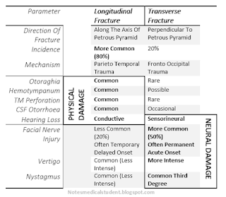

Parameter   Longitudinal Fracture Transverse Fracture   Direction Of  Fracture  Along The Axis Of Petrous Pyramid Perpendicular To Petrous Pyramid  Incidence  More Common (80%) 20%  Mechanism  Parieto Temporal Trauma Fronto Occipital Trauma  Otoraghia PHYSICAL DAMAGE Common Rare  Hemotympanum  Common Possible  TM Perforation  Common Rare  CSF Otorrhoea  Common Occasional  Hearing Loss  Conductive Sensorineural NEURAL DAMAGE Facial Nerve Injury   Less Common (20%) Often Temporary Delayed Onset More Common (50%) Often Permanent Acute Onset  Vertigo  Common (Less Intense) More Intense  Nystagmus  Common (Less Intense) Common Third Degree
Difference between temporal bone fracture types: Longitudinal Vs Transverse
Nerve Injury or neural component more common with transverse # Facial nerve injury(VII), vertigo(VIII: Vestibular Nerve), Sensorineural hearing loss(VIII: Auditory Nerve) and Nystagmus (vestibular Nerve/ CNS component) are more common and intense with transverse fracture.

Physical damage like bleeding demonstrated as  Hemotympanum and otorraghia , fractures seen as  CSF otorrhea, Conductive hearing loss due o disruption of ossicles and Tympanic membrane perforation, are common with longitudinal fracture. Neural component less common or less intense than it’s contrary.

Differential diagnoses of various otological symptoms

Parameter

Longitudinal Fracture
Transverse Fracture

Direction Of  Fracture
Along The Axis Of Petrous Pyramid
Perpendicular To Petrous Pyramid
Incidence
More Common (80%)
20%
Mechanism
Parieto Temporal Trauma
Fronto Occipital Trauma
Otoraghia
PHYSICAL DAMAGE
Common
Rare
Hemotympanum
Common
Possible
TM Perforation
Common
Rare
CSF Otorrhoea
Common
Occasional
Hearing Loss
Conductive
Sensorineural
NEURAL DAMAGE
Facial Nerve Injury

Less Common (20%)
Often Temporary
Delayed Onset
More Common (50%)
Often Permanent
Acute Onset
Vertigo
Common (Less Intense)
More Intense
Nystagmus
Common (Less Intense)
Common Third Degree


Sunday, September 17, 2017

Indications for Cesarean Section: Mnemonics


Cesarean Section is the delivery of the product of conception after the period of viability by making an incision in the anterior wall of the Uterus.

Cesarean section step by step procedure (Flowchart)


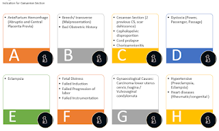
A  AntePartum Hemorrhage (Abruptio and Central Placenta Previa) B  Breech/ transverse (Malpresentation)  Bad Obstretric History  C  Cesarean Section (2 previous CS, scar dehiscence)  Cephalopelvic disproportion  Cord prolapse  Chorioamnionitis D  Dystocia (Power, Passenger, Passage)     E  Eclampsia F  Fetal Distress  Failed Induction  Failed Progression of labor   Failed Instrumentation G  Gynaecological Causes: Carcinoma lower uterus cervix /vagina / Vulvovaginal condylomata     H   Hypertensive (Preeclampsia, Eclampsia)  Heart diseases (Rheumatic/congenital )
Indication for Cesarean Section:  Mnemonic

Points to be noted are
1.       Product of Conception can be living or dead.
2.       Before viability, if POC is removed by undergoing surgical Procedure it will be called Hyste-RO-tomy (Not Hyste-REC-tomy) and not caesarean section.
3.       The delivery is made by making an incision on the anterior wall of the UTERUS. Though incision on anterior wall of the ABDOMEN is necessary to reach the anterior wall of uterus, the definition of cesarean section will be incomplete just by mentioning the anterior abdominal wall. So delivery of ruptured uterus and abdominal pregnancy is not C section.
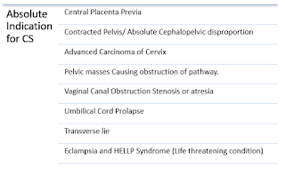
1. Central Placenta Previa 2. Contracted Pelvis/ Absolute Cephalopelvic disproportion 3. Advanced Carcinoma of Cervix 4. Pelvic masses Causing obstruction of pathway. 5. Vaginal Canal Obstruction Stenosis or atresia  6. Umbilical Cord Prolapse 7. Transverse lie 8. Eclampsia and HELLP Syndrome (Life threatening condition)
Absolute Indication For C Section

Absolute Indications for Cesarean section.

1.       Central Placenta Previa
2.       Contracted Pelvis/ Absolute Cephalopelvic disproportion
3.       Advanced Carcinoma of Cervix
4.       Pelvic masses Causing obstruction of pathway.
5.       Vaginal Canal Obstruction Stenosis or atresia
6.       Umbilical Cord Prolapse
7.       Transverse lie
8.       Eclampsia and HELLP Syndrome (Life threatening condition)
              
 Learn about history taking in Obstetrics.


Relative Indications

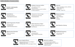
1. Relative Cephalopelvic Disproportion  2. Previous Cesarean section  a. Two previous C Section  b. Scar dehiscence c. Previous CS was for recurrent condition  d. Previous Classical CS 3. Dystocia  a. Power: Poor effort by mother, inefficient uterine contractions b. Passage: CPD c. Passenger:  Large Fetus,  4. Fetal Distress  a. Non Reassuring CTG  b. Fetal Asphyxia and acidosis 5. Antepartum Hemorrhage a. Placenta Previa (type IIb,III) b. Abruptioplacentae 6. Malpresentation  a. Primi Breech  b. Non Frank Breech  c. Brow  d. Shoulder 7. Failed Progression of Labor a. Secondary Arrest  b. Prolonged Labor  c. Failed Instrumental Delivery  8. Failed Induction of labor (Drugs, Artificial Rupture of Membrane) 9. Bad Obstetrics history  10. Hypertensive Disorders a. Severe Preeclampsia b. Eclampsia 11. Infections  a. Chorioamnionitis b. HPV (Condyloma Acuminata) c. HIV  12. Multifetal Pregnancy  a. Mono amniotic twin  b. Conjoint Twins  13. Gynecological Illness a. Mechanical Obstruction due to Benign or Malignant pelvic tumor  b. Following repair of VVF (vesicovaginal Fistula) 14. Medical Conditions a. Uncontrolled Diabetes Mellitus  b. Heart Disease (Rheumatic/ Congenital/ Coarctation of aorta) c. Marfan Syndrome
Relative Indications of CS

1.       Relative Cephalopelvic Disproportion
2.       Previous Cesarean section
a.       Two previous C Section
b.      Scar dehiscence
c.       Previous CS was for recurrent condition
d.      Previous Classical CS
3.       Dystocia
a.       Power: Poor effort by mother, inefficient uterine contractions
b.      Passage: CPD
c.       Passenger:  Large Fetus,
4.       Fetal Distress
a.       Non Reassuring CTG
b.      Fetal Asphyxia and acidosis
5.       Antepartum Hemorrhage
a.       Placenta Previa (type IIb,III)
b.      Abruptioplacentae
6.       Malpresentation
a.       Primi Breech
b.      Non Frank Breech
c.       Brow
d.      Shoulder
7.       Failed Progression of Labor
a.       Secondary Arrest
b.      Prolonged Labor
c.       Failed Instrumental Delivery
8.       Failed Induction of labor (Drugs, Artificial Rupture of Membrane)
9.       Bad Obstetrics history
10.   Hypertensive Disorders
a.       Severe Preeclampsia
b.      Eclampsia
11.   Infections
a.       Chorioamnionitis
b.      HPV (Condyloma Acuminata)
c.       HIV
12.   Multifetal Pregnancy
a.       Mono amniotic twin
b.      Conjoint Twins
13.   Gynecological Illness
a.       Mechanical Obstruction due to Benign or Malignant pelvic tumor
b.      Following repair of VVF (vesicovaginal Fistula)
14.   Medical Conditions
a.       Uncontrolled Diabetes Mellitus
b.      Heart Disease (Rheumatic/ Congenital/ Coarctation of aorta)
c.       Marfan Syndrome


Learn about Gravida, Para with clinical case scenarios.


Thursday, August 31, 2017

Mnemonics: Breast Cancer causes

Risk Factors for Development of Breast Cancer (A to G Guide)

For easy remembrance of the risk factors that can cause breast cancer, it is grouped into 7 major titles that start from A to G, and under each heading both protective factors or factors that has least causative effect and factors that has strong risk are classified and described.
Risk Factors for development of Breast Carcinoma


Risk
Protective
A
Age
Old age
>90 years (~20%)
Rare <20 years
B
Benign Breast Conditions
Hyperplasia (1.5-3 fold)
Papilloma (1.5-3 fold)
Ductal / Lobular Atypical hyperplasia (5 fold)

C
Cancer
Cancer of contralateral Breast (3-4 fold)
Cervix
Ovary
Colon
Prostate
Radiation Therapy for Hodgkin's Lymphoma

D
Diet
Alcohol
Phytoestrogen Deficiency
Vitamin C
E
Estrogen
(Conversion of Steroid hormone to estradiol in the body fat)
Nulliparous
Obese Postmenopausal Women
Exogenous Estrogen (OCP, Mixed prep HRT)
Breast feeding
Late Menarche
Early Menopause
Early age at 1st child birth
F
Familial History
2-10 fold risk in family history of Breast, Colon, Ovarian and Prostate Carcinoma

G
Genetics
BRCA 1 (Breast Ovarian, prostate and Colorectal Ca )
BRCA2 (Male Breast Ca)
p53
HER 2

G
Geography
African american
(Western world 1-3 % of all death of women )
Rare in Japan and Taiwan
Source: Bailey and Love’s Short Practice of Surgery.

Tuesday, August 1, 2017

Causes of altered sensation of Smell


Anosmia (No smell)
Atrophic rhinitis
Peripheral neuritis
Degenerative Disease of Nose
Injury to factory nerve
Injury to olfactory bulb
Anterior Cranial fossa fracture
Intracranial abscess
Intracranial tumors
Meningitis

Causes of various types of Nasal discharge.

Parosmia (Perversion of smell)
Recovery Phase of post influenza anosmia
Intracranial tumor

Clinical Examination of Nose and PNS

Hyposmia (Decreased Smell)
Nasal Polyp
Enlarged Turbinate
Edema of Mucosal Membrane
 Common cold
Vasomotor Rhinitis
Allergic Rhinitis


Wednesday, July 5, 2017

Rule of 9: Burn

Wallace Rule of Nine
For rough estimation of the body surface area that is affected by Burn, Wallace rule of nine is used. It  is used for adult. Slight modification is done in case of children as they have larger head compared to adults and hence Lund and Browder chart is used. The skin is grossly allocated the value of 9% as demostrated in the chart and diagram below.

Source: https://twitter.com/pjonline_news/status/596994284466802688
Wallace Rule of Nine
(source: https://twitter.com/pjonline_news/status/596994284466802688)

Body part

Head
9%
Right upper extremity
9%
Left Upper extremity
9%
Anterior Chest
9%
Posterior Chest
9%
Anterior right lower extremity
9%
Posterior left lower extremity
9%
Posterior right lower extremity
9%
Posterior left lower extremity
9%
Perineum
1%
Total
100%


Friday, June 23, 2017

Rule of 10 : Pheochromocytoma

As previous site was dedicated to certain medical conditions that can be described with a certain number. So is this blog. This collects Number 5-10.

Rule of 10

Pheochromocytoma 

Pheochromocytoma is a tumor most commonly arising from the adrenal medulla and secrete catecholamines (Adrenaline and Noradrenaline) in the vascular system however 10% arise from extraadrenal sites such as sympathetic chain around spinal cord, aorta, ureter and bladder. Generally these are benign tumors but 10% are malignant. 90 % occurs unilaterally and 10% occur in the family. Recently 10 gene mutations have been associated with the condition to add up to its list of 10. It rarely occurs in children (10%). And has tendency to recur (10%) even after surgical excision. 10% have been identified after the patient develops Stroke following malignant Hypertension.

Pheochromocytoma
Rule of 10 in pheochromocytoma

Monday, February 27, 2017

Pupillary Reaction to Various Drugs and Substances

PUPIL REACTION TO SUBSTANCE/DRUGS

Pupil of human eye constrict and dilate depending upon the need of the eye to focus on near object/far object, dim light/bright light and whether it is under stress/not. This response is solely dependent upon the muscles inside the eyes in the iris. The pupillae constrictor muscle respond to cholinergic agents and cause pupillary constriction while the dilator pupillae muscle is acted upon by sympathetic system and causes pupillary dilation.

Beside these natural response, the substances we come in contact with can also alter the pupillary size. Drugs such as Adrenaline, antidepresssants, antihistaminics, can cause pupillary dilatation. Pethidine being an exception is the only opoid to cause pupillary dilation. Herbal products such as belladona, strychinine, calotropis and Chemicals such as alcohol, formic acid, cyanide Carbon monoxide also have dilatatory effect based upon the dosing.

Phenols, Organophosphate, carbamates, Carbolic acid, cause pupillary cosntriction. Drugs such as Opoids, barbiturates, Benzodiaepines, methyl dopa and plant product such as Nicotine, caffiene and opium causes the dilatation of pupil. 
DILATORS 
CONSTRICTORS
A6 C5 (Big1 Co Ca Cola Can)
O2 B2 C5 ( Car Car Caf )
Formic Acid


Phenol (Carbolic Acid)
Pethidine2 (only exception in Opoid)

O
All Opoids2  (Except severe Acidosis/hypoxia/ respiratory depression)
Adrenaline*/ Ephidrine/Sympathetomimetics
A

Parasympathetomimetics*
Atropine*/ Belladona/ Dhatura
A
O
Organophosphate*
Amphetamine
A
B
Barbiturates
Alcohol3
A
B
Benzodiazepines
Antidepressants
A


Antihistaminics
A


Cocaine Crack
Co
Car
Carbamate
Carbon Monoxide/ CO
Ca/CO
Car
Carbolic Acid
Calotropis

Cola
Caf
Caffeine
Cannabis
Can

Coma of Alcohol3
Cyanide


Clonidine


Hypothermia*


Heat Stroke*
Strychnine


Nicotine4


Methyl Dopa
HIPPUS: Alternating dilatation and constriction is done by ACONITE
Note: All A’s constrict the pupil. All B’s and O’s dilate the pupil.
1 Big is not a part of drug rather means dilatation.
2 Only opoid to cause pupillary dilatation is Pethidine.
3 Toxic Effect of alcohol causes pupillary constriction as patient develops coma.
4 Delayed effect of Nicotine is dilatation of pupil.
* The drugs/substances in row with different color across the table are physiological Counterparts